Bacteria are a large group of minute, unicellular, microscopic organisms, which have been classified as prokaryotic cells, as they lack a true nucleus. These microscopic organisms comprise a simple physical structure, including cell wall, capsule, DNA, pili, flagellum, cytoplasm and ribosomes.
Bacteria can be gram-positive or gram-negative depending upon the staining methods. Let us have a detailed look at the difference between the two types of bacteria.
Gram Staining
This technique was proposed by Christian Gram to distinguish the two types of bacteria based on the difference in their cell wall structures. The gram-positive bacteria retain the crystal violet dye, which is because of their thick layer of peptidoglycan in the cell wall.
This process distinguishes bacteria by identifying peptidoglycan that is found in the cell wall of the gram-positive bacteria. A very small layer of peptidoglycan is dissolved in gram-negative bacteria when alcohol is added.
Gram-Positive and Gram-Negative Bacteria – Overview
The gram-positive bacteria retain the crystal violet colour and stain purple whereas the gram-negative bacteria lose crystal violet and stain red. Thus, the two types of bacteria are distinguished by gram staining.
Gram-negative bacteria are more resistant to antibodies because their cell wall is impenetrable.
Gram-positive and gram-negative bacteria are classified based on their ability to hold the gram stain. The gram-negative bacteria are stained by a counterstain such as safranin, and they are de-stained because of the alcohol wash. Hence under a microscope, they are noticeably pink in colour. Gram-positive bacteria, on the other hand, retains the gram stain and show a visible violet colour upon the application of mordant (iodine) and ethanol (alcohol).
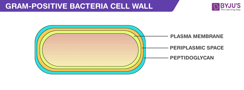
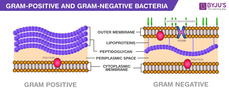
Gram-positive bacteria constitute a cell wall, which is mainly composed of multiple layers of peptidoglycan that forms a rigid and thick structure. Its cell wall additionally has teichoic acids and phosphate. The teichoic acids present in the gram-positive bacteria are of two types – the lipoteichoic acid and the teichoic wall acid.
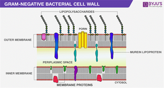
In gram-negative bacteria, the cell wall is made up of an outer membrane and several layers of peptidoglycan. The outer membrane is composed of lipoproteins, phospholipids, and LPS. The peptidoglycan stays intact to lipoproteins of the outer membrane that is located in the fluid-like periplasm between the plasma membrane and the outer membrane. The periplasm is contained with proteins and degrading enzymes which assist in transporting molecules.
The cell walls of the gram-negative bacteria, unlike the gram-positive, lacks the teichoic acid. Due to the presence of porins, the outer membrane is permeable to nutrition, water, food, iron, etc.
Gram Positive Bacteria
Introduction
Gram-positive bacteria are the genus of the bacteria family and a member of the phylum Firmicutes. These bacteria retain the colour of the crystal violet stain which is used during gram staining. These bacteria give a positive result in the Gram stain test by appearing purple coloured when examined under a microscope, hence named, gram-positive bacteria. Actinomyces, Clostridium, Mycobacterium, Streptococci, Staphylococci, and Nocardia are a few examples of Gram-positive bacteria.
Gram-positive bacteria are the genus of the bacteria family and a member of the phylum Firmicutes. These bacteria retain the colour of the crystal violet stain which is used during gram staining. These bacteria give a positive result in the Gram stain test by appearing purple coloured when examined under a microscope, hence named, gram-positive bacteria. Actinomyces, Clostridium, Mycobacterium, Streptococci, Staphylococci, and Nocardia are a few examples of Gram-positive bacteria.
Characteristics of Gram-Positive Bacteria
- They have a thick peptidoglycan layer and cytoplasmic lipid membrane.
- These bacteria lack an outer membrane.
- Have a lower lipid content and more teichoic acids.
- They move around with the help of locomotion organs such as cilia and flagella.
- The walls of Staphylococcus aureus and Streptococcus faecalis contain teichoic acid.
Gram-Positive Cell Wall


Peptidoglycan
It is a permeable, cross-linked organic polymer and rigid structure which plays an important role in providing shape and strength to the cell wall. It makes up about 90% of the cell wall enclosing the plasma membrane and protects the cell from the environment. Peptidoglycan is composed of three main components including Glycan backbone, Peptide, and Tetra-peptide.
It is a permeable, cross-linked organic polymer and rigid structure which plays an important role in providing shape and strength to the cell wall. It makes up about 90% of the cell wall enclosing the plasma membrane and protects the cell from the environment. Peptidoglycan is composed of three main components including Glycan backbone, Peptide, and Tetra-peptide.
Lipid
The lipid element found in the gram-positive bacteria cell wall supports its anchoring to the membrane. The total percentage of lipid content in a gram-positive bacterium cell wall is 2 – 5 per cent.
The lipid element found in the gram-positive bacteria cell wall supports its anchoring to the membrane. The total percentage of lipid content in a gram-positive bacterium cell wall is 2 – 5 per cent.
Teichoic acid
It is water-soluble and a polymer of glycerol. Teichoic acid is the major surface antigen of gram-positive bacteria and it makes up about fifty per cent of the total dry weight of the cell wall.
It is water-soluble and a polymer of glycerol. Teichoic acid is the major surface antigen of gram-positive bacteria and it makes up about fifty per cent of the total dry weight of the cell wall.
Benefits of Gram-Positive Bacteria
- These species of bacteria are non-pathogenic and reside within our body including the mouth, skin, intestine, and upper respiratory tract.
- They are an essential ingredient in producing Emmentaler or Swiss cheese.
- The species of Corynebacterium are used in the industrial production of enzymes, amino acids, nucleotides, etc.
- Various Bacillus species are used in the secretion of large quantities of enzymes.
- A few species of gram-positive bacteria are also involved in cheese ageing, bioconversion of steroids, degradation of hydrocarbons, etc.
- Bacillus amyloliquefaciens of gram-positive bacteria are a good source of a natural antibiotic protein – Barnase.
Risk Factors of Gram-Positive Bacteria
The substantial increase in skin and mucous infections in all humans are caused by staphylococcal species. These organisms are mainly transmitted through skin contact, fomite contact, inhaling infected aerosolized particles, by pets, etc. Other risk factors include food poisoning, Respiratory Diseases, tooth cavities, Diphtheria, Mycobacterium tuberculosis, etc.
The substantial increase in skin and mucous infections in all humans are caused by staphylococcal species. These organisms are mainly transmitted through skin contact, fomite contact, inhaling infected aerosolized particles, by pets, etc. Other risk factors include food poisoning, Respiratory Diseases, tooth cavities, Diphtheria, Mycobacterium tuberculosis, etc.
Gram Negative Bacteria
Gram negative bacteria are the genus of bacteria family and a member of the phylum Firmicutes. They are the group of aerobic bacteria which does not retain the crystal violet dye during the procedure of Gram staining and appear pink in colour when examined under the microscope.
There are several gram negative bacteria with medical significance. The most important of these are members of the family Enterobacteriaceae. Further genera of Gram negative bacteria include Vibrio, Campylobacter, Pseudomonas, and other bacteria which are normally found in the gastrointestinal tract.
General Characteristics of Gram Negative Bacteria
The gram negative bacteria have the following characteristics:
- The cell wall is thin without an outer layer.
- A high percentage of lipids can be found.
- It contains all types of amino acids.
- The muramic acid content is less.
- It is sensitive to streptomycin.
- It is devoid of magnesium ribonucleate and teichoic acid.
- It contains lipopolysaccharides, sialic acid, and flagella.
Cell Structure of Gram Negative Bacteria

- The cell wall of Gram negative bacteria is thin and is composed of peptidoglycan.
- The cell envelope has 3 layers including, a unique outer membrane, a thin peptidoglycan layer, and the cytoplasmic membrane.
- An outer membrane of the cell wall is a bilayer structure consisting of phospholipids molecules, lipopolysaccharides (LPS), lipoproteins and surface proteins.
- Endotoxin is toxins released by the cell during infections and function as receptors and blocking immune response.
- The porin proteins are present in the upper layer of a cell which functions by regulating the entry and exit of the molecules within the cell.
Diseases caused by Gram Negative Bacteria
The diseases caused by gram negative bacteria are diarrhoea, inflammatory disease of the large intestine, infantile diarrhoea, kidney damage, typhoid fever, bubonic plague, cholera etc.
The diseases caused by gram negative bacteria are diarrhoea, inflammatory disease of the large intestine, infantile diarrhoea, kidney damage, typhoid fever, bubonic plague, cholera etc.
Difference between Gram-Positive and Gram-Negative Bacteria – Key Points
- The cell wall of gram-positive bacteria is composed of thick layers peptidoglycan.
- The cell wall of gram-negative bacteria is composed of thin layers of peptidoglycan.
- In the gram staining procedure, gram-positive cells retain the purple coloured stain.
- In the gram staining procedure, gram-negative cells do not retain the purple coloured stain.
- Gram-positive bacteria produce exotoxins.
- Gram-negative bacteria produce endotoxins.
Difference between Gram-Positive and Gram-Negative Bacteria
Following are the important differences between gram-positive and gram-negative bacteria:

Difference between Gram-Positive and Gram-Negative Bacteria
| Gram-Positive bacteria | Gram-Negative bacteria |
| Cell Wall | |
| A single-layered, smooth cell wall | A double-layered, wavy cell-wall |
| Cell Wall thickness | |
| The thickness of the cell wall is 20 to 80 nanometres | The thickness of the cell wall is 8 to 10 nanometres |
| Peptidoglycan Layer | |
| It is a thick layer/ also can be multilayered | It is a thin layer/ often single-layered. |
| Teichoic acids | |
| Presence of teichoic acids | Absence of teichoic acids |
| Outer membrane | |
| The outer membrane is absent | The outer membrane is present (mostly) |
| Porins | |
| Absent | Occurs in Outer Membrane |
| Mesosome | |
| It is more prominent. | It is less prominent. |
| Morphology | |
| Cocci or spore-forming rods | Non-spore forming rods. |
| Flagella Structure | |
| Two rings in basal body | Four rings in basal body |
| Lipid content | |
| Very low | 20 to 30% |
| Lipopolysaccharide | |
| Absent | Present |
| Toxin Produced | |
| Exotoxins | Endotoxins or Exotoxins |
| Resistance to Antibiotic | |
| More susceptible | More resistant |
| Examples | |
| Staphylococcus, Streptococcus, etc. | Escherichia, Salmonella, etc. |
| Gram Staining | |
| These bacteria retain the crystal violet colour even after they are washed with acetone or alcohol and appear as purple-coloured when examined under the microscope after gram staining. | These bacteria do not retain the stain colour even after they are washed with acetone or alcohol and appear as pink-coloured when examined under the microscope after gram staining. |


0 Comments