Human Eye, a precious gift from nature. Our eyes are the windows to the wonderful world, all we know and love, experience and discover, ponder and cherish. You can identify a flower by smelling and touching it, but you can’t identify the color without the sense of vision. Therefore vision is the most important sense organ out of five senses. Let’s peer into the structure and working of Human Eye.
Structure of Human Eye
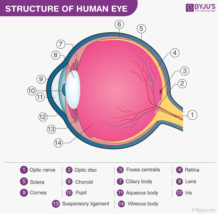
Nature has located eyes close to the brain so that its messages may be arrived there quickly. Eyeball owns a spherical shape and is completely surrounded by a layer of soft fatal tissue and placed within a bony orbit where eye is protected. A white colored stalk is known as Optic Nerve which connects the eye to the brain and also to the muscles which moves the eyeball.
Structure of eye seems to have three layers –
The first and outermost layer is known as Sclera which serves to protect the delicate structure within. The transparent bulging out portion is called Cornea and also crystalline lens which is one of the main parts of the human eye.
The second layer is called choroid which consists of three different zones-choroid proper that carries nourishment to the tissues of the eye. Another zone is called the ciliary body, a broad ring shaped band of thin muscles fiber which plays a very important role in vision adjustment of the eye. The third zone is called as Iris which expands and contracts the pupil much like the wi-frame of the camera.
The Third layer is called as Retina, a part of the optic nerve which transmit light impulses to the brain. It is the most important and complex structure in the eyeball, consists of complicated arrangements of rods and cones which convert light waves into nerve impulses.
Between the lense and the cornea is the Aqueous humor which consists of water and salt. The largest part in eye is called as Vitreous humor, consists of water mixed with salt and albumin. It is a highly transparent jelly like substance and plays an important role in visual adjustment.
The reflected light from outside enters through the crystal transparency of the cornea, pupil, Aqueous humor, lens and vitreous humor which is a clear gel-like substance that fills the middle of the eye and then projects on to the photoreceptor of the Retina, which is a photo sensitive tissue lying at the back of the eye.
The macula is a very small area at the centre of the Retina that gives a fine pin point centre of vision. The area of the Retina surrounding the macula gives a peripheral or sight vision. The retina convert the light rays into the signals that are sent to the optic nerve and then to the brain. Continuous adjustment of the pupil and lens regulates the entry and focusing light.
Structure of Human Eye
A human eye is roughly 2.3 cm in diameter and is almost a spherical ball filled with some fluid. It consists of the following parts:

- Sclera: It is the outer covering, a protective tough white layer called the sclera (white part of the eye).
- Cornea: The front transparent part of the sclera is called the cornea. Light enters the eye through the cornea.
- Iris: A dark muscular tissue and ring-like structure behind the cornea is known as the iris. The colour of the iris actually indicates the colour of the eye. The iris also helps regulate or adjust exposure by adjusting the iris.
- Pupil: A small opening in the iris is known as a pupil. Its size is controlled with the help of iris. It controls the amount of light that enters the eye.
- Lens: Behind the pupil, there is a transparent structure called a lens. By the action of ciliary muscles, it changes its shape to focus light on the retina. It becomes thinner to focus on distant objects and becomes thicker to focus on the nearby objects.
- Retina: It is a light-sensitive layer that consists of numerous nerve cells. It converts images formed by the lens into electrical impulses. These electrical impulses are then transmitted to the brain through optic nerves.
- Optic nerves: Optic nerves are of two types. These include cones and rods.
- Cones: Cones are the nerve cells that are more sensitive to bright light. They help in detailed central and colour vision.
- Rods: Rods are the optic nerve cells that are more sensitive to dim lights. They help in peripheral vision.
At the junction of the optic nerve and retina, there are no sensory nerve cells. So no vision is possible at that point and is known as a blind spot.
An eye also consists of six muscles. It includes the medial rectus, lateral rectus, superior rectus, inferior rectus, inferior oblique, and superior oblique. The basic function of these muscles is to provide different tensions and torques that further control the movement of the eye.
Function of the Human Eye
As we mentioned earlier, the eye of a human being is like a camera. Much like the electronic device, the human eye also focuses and lets in light to produce images. So basically, light rays that are deflected from or by distant objects land on the retina after they pass through various mediums like the cornea, crystalline lens, aqueous humor, the lens, and vitreous humor.

The concept here though is that as the light rays move through the various mediums, they experience refraction of light. Well, to put it in simple terms, refraction is nothing but the change in direction of the rays of light as they pass between different mediums. The table below shows the refractive indices of the various parts of the eye.

Having different refractive indexes is what bends the rays to form an image. The light rays finally are received and focused on the retina. The retina contains photoreceptor cells called rods and cones and these basically detect the intensity and the frequency of the light. Further, the image that is formed is processed by millions of these cells, and they also relay the signal or nerve impulses to the brain via the optic nerve. The image formed is usually inverted but the brain corrects this phenomenon. This process is also similar to that of a convex lens.
In any case, now that we have learned something about the human eye, each eye is very important, and they play a distinct part in helping humans to see.
As we mentioned earlier, the eye of a human being is like a camera. Much like the electronic device, the human eye also focuses and lets in light to produce images. So basically, light rays that are deflected from or by distant objects land on the retina after they pass through various mediums like the cornea, crystalline lens, aqueous humor, the lens, and vitreous humor.

The concept here though is that as the light rays move through the various mediums, they experience refraction of light. Well, to put it in simple terms, refraction is nothing but the change in direction of the rays of light as they pass between different mediums. The table below shows the refractive indices of the various parts of the eye.

Having different refractive indexes is what bends the rays to form an image. The light rays finally are received and focused on the retina. The retina contains photoreceptor cells called rods and cones and these basically detect the intensity and the frequency of the light. Further, the image that is formed is processed by millions of these cells, and they also relay the signal or nerve impulses to the brain via the optic nerve. The image formed is usually inverted but the brain corrects this phenomenon. This process is also similar to that of a convex lens.
In any case, now that we have learned something about the human eye, each eye is very important, and they play a distinct part in helping humans to see.
Human Eye
The human eyes are the most complex sense organ in the human body, which comprises several parts to form a spherical structure. The structure of the human eye can be primarily classified into external structure and internal structure. Every aspect of the human eye is mainly responsible for a specific action.
Parts of an eye
Based on their visibility and their location, parts have been classified into:
The External Structure of an Eye
The parts of the eye that are present outside and are visible externally comprise the eye’s external structure. The external or outer structure of the eye includes the following parts:
- Iris
- Pupil
- Sclera
- Cornea
- Conjunctiva
The Internal Structure of an Eye
The parts of the eye enclosed in a socket, which is composed of several of the bones of the skull, are called the internal structure of the eye. The internal structure of the eye includes the following parts:
- Lens
- Retina
- Optic nerve
- Vitreous Humour
- Aqueous Humour
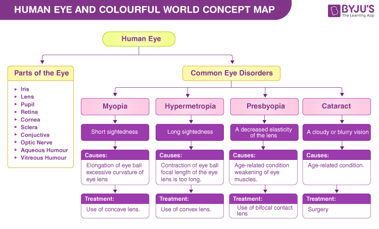
The human eye concept map describes essential parts of an eye and a few common eye disorders.

Defects of a Human Eye
There are few common eye disorders seen in all individuals, and they are caused by several factors. These conditions can be improved by the corrections. The defects include:
Myopia – This is also called short-sightedness. A person with this eye defect can only see nearby objects clearly compared to distant objects. This condition can be corrected using a concave lens.

Hypermetropia – This is also called farsightedness. A person with this eye defect can only see distant objects clearly compared to near objects. This condition can be corrected using a convex lens.

Presbyopia – This is an age-related condition caused due to the weakening of ciliary muscles, hardening of the lens, and reduced lens flexibility. A person with this defect usually finds difficulties focusing on nearby objects and is unable to read or write.
Cataract – This is an age-related condition caused due to the loss of transparency of the lens by erosion of lens proteins. It usually results in blurry vision and cloudy lenses and can be corrected by replacing the old lens with an artificial lens.


0 Comments